
🩺 INTRODUCTION
Every successful cardiac operation begins long before the first incision. Increasingly, its foundation is built not in the operating room, but in the imaging suite.
For generations, cardiac surgery relied on clinical expertise, physical examination, conventional angiography, and the surgeon’s experience to guide complex decisions. While these remain indispensable, today’s cardiac surgeon has access to something previous generations could only imagine: an extraordinarily detailed, three-dimensional understanding of the heart before surgery even begins.
Modern cardiac imaging has transformed how we diagnose disease, assess risk, plan operations, and anticipate challenges. It has shifted cardiac surgery from reacting to disease to preparing for it with unprecedented precision.
Today, imaging is no longer simply diagnostic. It has become an essential partner in surgical decision-making.
🔍 MORE THAN JUST PICTURES
Many patients think of CT scans or MRIs as tests that simply “take images.”
In reality, advanced cardiac imaging provides a comprehensive roadmap of the heart’s anatomy, function, blood flow, tissue characteristics, and structural integrity.
Rather than answering “Is something wrong?”, modern imaging helps answer far more important clinical questions:
- What exactly is the disease?
- How severe is it?
- Which treatment offers the greatest long-term benefit?
- Can surgery be performed more safely?
- What challenges should be anticipated before entering the operating room?
These answers fundamentally influence how cardiac surgeons plan each operation.
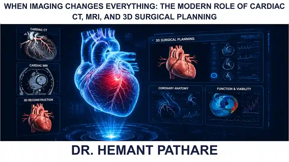
🖥️ CARDIAC CT: MAPPING THE CORONARY LANDSCAPE
Cardiac Computed Tomography (CT) has evolved into one of the most valuable tools in cardiovascular medicine.
Beyond identifying coronary artery disease, modern CT imaging provides exceptional visualization of coronary anatomy, calcification, bypass grafts, cardiac chambers, and the thoracic aorta.
For surgeons, CT frequently reveals details that influence operative planning, including the extent of vascular calcification, anatomical variations, previous surgical changes, and the relationship of vital structures. In many complex cases, knowing these details beforehand significantly improves procedural safety and efficiency.
Outpatient-based MDCT coronary angiography is fast replacing in-hospital conventional angiography as the preferred means of elective imaging of the coronaries unless there is significant calcification, wherein a calcium score lets us know that conventional angiography would be better. In non-cardiac surgical cases, in fact, it is the go-to investigation when there is suspicion of age-related coronary artery disease.
🧲 CARDIAC MRI: UNDERSTANDING THE HEART BEYOND ANATOMY
While CT excels in anatomical detail, Cardiac Magnetic Resonance Imaging (MRI) offers something equally valuable: a detailed understanding of the heart muscle itself.
Cardiac MRI can evaluate:
- Myocardial viability
- Percentage of scar tissue after myocardial infarction
- Cardiomyopathies
- Inflammatory heart diseases
- Ventricular function
- Valvular heart disease
This information helps determine not only whether surgery should be performed, but also when it is likely to provide the greatest benefit.
Sometimes the most important surgical decision is recognizing when an operation will meaningfully improve both survival and quality of life.
🧩 THE RISE OF THREE-DIMENSIONAL SURGICAL PLANNING
Perhaps one of the most exciting developments in modern cardiac surgery is the use of three-dimensional reconstruction.
Rather than interpreting multiple two-dimensional images, surgeons can now visualize complex cardiac anatomy in three dimensions before entering the operating room.
For congenital heart disease, structural valve abnormalities, complex aortic pathology, and redo cardiac operations, 3D planning offers an invaluable opportunity to anticipate anatomical challenges and refine surgical strategy.
Every detail studied beforehand is one less surprise encountered during surgery.
🤖 IMAGING MEETS ARTIFICIAL INTELLIGENCE
Artificial Intelligence is rapidly becoming an important extension of advanced cardiac imaging.
AI-assisted algorithms can now support image reconstruction, automate anatomical measurements, identify subtle abnormalities, and improve workflow efficiency.
While these technologies are not replacing clinical expertise, they are enhancing diagnostic accuracy and enabling clinicians to make faster, more informed decisions.
The future of imaging will likely combine machine intelligence with human judgment, creating a partnership that benefits both surgeons and patients.
🎯 PRECISION BEGINS BEFORE THE FIRST INCISION
The greatest strength of modern imaging is not simply producing clearer images.
It is enabling personalized care.
Every patient’s anatomy is different. Every coronary artery follows its own course. Every valve, every chamber, and every surgical challenge is unique.
Advanced imaging allows cardiac surgeons to tailor each procedure to the individual rather than applying a standardized approach to every case.
This is precision medicine in practice.
🚀 LOOKING AHEAD
Cardiac imaging continues to evolve at an extraordinary pace. Hybrid OTs with built-in imaging modalities are a boon for transfemoral or transcarotid aortic valve replacements in the geriatric population. Likewise, they are invaluable for device closure of atrial septal defects.
Emerging technologies such as photon-counting CT, four-dimensional flow MRI, AI-assisted image interpretation, virtual reality visualization, and patient-specific digital heart models are expected to further transform surgical planning in the coming years.
The future is not merely about seeing the heart more clearly.
It is about understanding it more completely.
As imaging becomes increasingly sophisticated, cardiac surgery will continue to become safer, more personalized, and more predictable.
💡 FINAL THOUGHT
Every remarkable operation begins with remarkable preparation.
Modern cardiac imaging has changed the way we approach surgery, allowing us to anticipate complexity rather than react to it. It empowers clinicians to make better-informed decisions, reduce uncertainty, and deliver care that is increasingly tailored to the individual patient.
The operating room will always remain the stage where skill and experience come together.
But the success of many cardiac procedures is now determined long before the patient enters it, through the clarity, precision, and insight provided by advanced imaging.
In many ways, the future of cardiac surgery will not simply be defined by better operations, but by better planning.
❓ A QUESTION WORTH REFLECTING ON
If advanced imaging can reveal the heart in extraordinary detail before surgery even begins, how might the next generation of technologies transform not only how we operate, but how we prevent, personalize, and predict cardiovascular disease?
Perhaps the greatest innovation in cardiac surgery is not learning to operate differently, but learning to understand every heart more completely before the first incision is ever made.
