
🩺 INTRODUCTION
Not all coronary artery disease is the same. While some blockages are soft and relatively straightforward to treat, others become hardened by years of calcium deposition, transforming routine procedures into some of the most technically demanding challenges in cardiovascular medicine.
Coronary artery calcification is far more than an imaging finding. It is a marker of advanced atherosclerosis, chronic vascular inflammation, and increasing procedural complexity.
For cardiac surgeons and interventional cardiologists alike, heavily calcified coronary arteries demand not only technical expertise but also careful planning, multidisciplinary decision-making, and individualized treatment strategies.
In modern cardiac surgery, one of the greatest challenges is often not the blockage itself, but the calcium hidden within it.
🧱 WHAT IS CORONARY ARTERY CALCIFICATION?
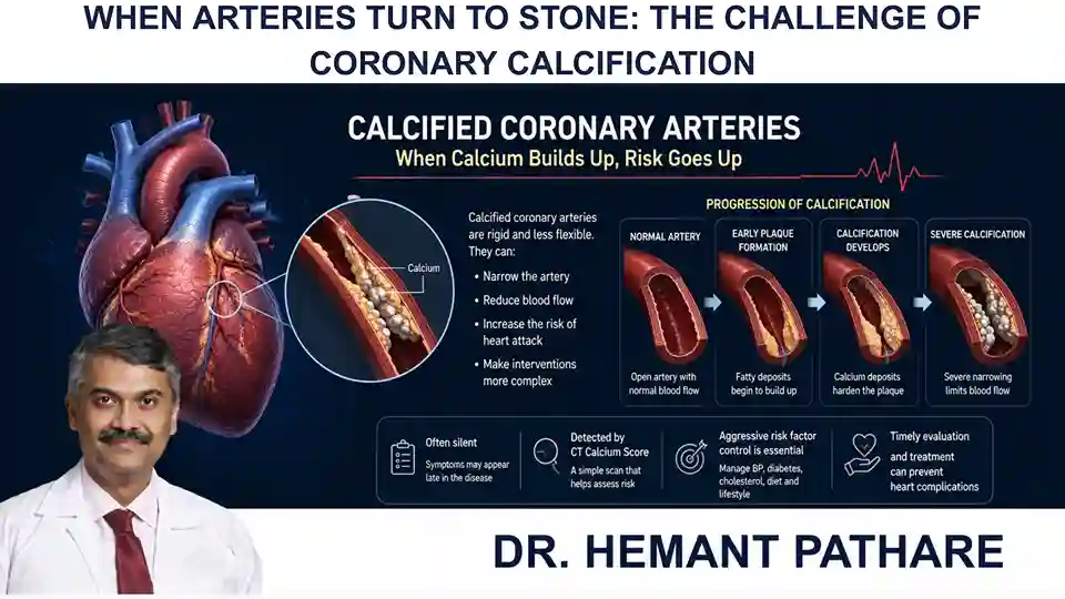
Coronary artery calcification (CAC) occurs when calcium deposits accumulate within the walls of the coronary arteries over many years as part of the atherosclerotic process.
Contrary to popular belief, calcium does not directly “cause” coronary artery disease. Rather, it reflects the body’s long-term response to chronic inflammation, lipid accumulation, and vascular injury.
The result is a vessel that becomes progressively rigid, less compliant, and increasingly difficult to treat.
Calcification is more commonly seen in older adults, individuals with diabetes, chronic kidney disease, hypertension, familial hypercholesterolemia, and long-standing cardiovascular disease.
⚠️ WHY CALCIUM CHANGES EVERYTHING
A calcified artery behaves very differently from a non-calcified one.
Its rigid structure limits vessel expansion, complicates bypass grafting and coronary interventions, and increases the technical complexity of both surgical and catheter-based procedures.
In severe cases, dense calcification can make coronary arteries difficult to manage. During Percutaneous Coronary Intervention (PCI), a diamond-tipped burr or rotablator is required to negotiate the narrowed calcified artery (with its attendant risks of perforation) before placing a balloon-expandable stent (while also increasing the risk of incomplete stent expansion).
During CABG surgery, it becomes difficult to cut into or graft the vessel since even the CC needles do not pass through the vessel wall and often a more laborious and unpredictable endarterectomy is required (which may result in a snow-ploughing effect on the microvasculature).
What appears to be a single coronary blockage on angiography may, in reality, represent a far more challenging anatomical problem. Understanding the extent and distribution of calcium before treatment is therefore critical to achieving durable outcomes.
🖥️ SEEING WHAT CONVENTIONAL ANGIOGRAPHY CANNOT
Modern cardiac imaging has fundamentally changed how coronary calcification is evaluated.
Coronary CT angiography and dedicated coronary calcium scoring provide valuable insight into both the burden and distribution of calcified plaque before any intervention is planned. However, if there is significant calcium scoring, then a CT coronary angiogram cannot be carried out, and a conventional coronary angiogram is required.
Intravascular Ultrasound (IVUS) and Optical Coherence Tomography (OCT) further allow cardiologists and clinicians to assess plaque morphology, calcium thickness, and lesion characteristics from within the artery itself.
These imaging technologies have shifted decision-making from estimation to precision, enabling clinicians to select the safest and most effective treatment strategy for each patient.
🎯 CHOOSING THE RIGHT STRATEGY
There is no universal solution for heavily calcified coronary disease. Statins themselves result in earlier calcification of soft plaques. Paradoxically, I find performing a coronary endarterectomy on a beating heart is easier than after the heart has been arrested.
Treatment depends on multiple factors, including coronary anatomy, lesion complexity, ventricular function, patient comorbidities, and overall surgical risk.
For many patients with diffuse multivessel calcification, Coronary Artery Bypass Grafting (CABG) continues to provide durable long-term revascularization.
In other cases, advances in PCI, including rotational atherectomy, orbital atherectomy, intravascular lithotripsy, and specialized balloon technologies, have expanded the options available for managing complex calcified lesions.
The question is no longer whether calcification can be treated, but which approach offers the greatest long-term benefit for the individual patient.
🚀 INNOVATION IS CHANGING THE LANDSCAPE
Until recently, severe coronary calcification often limited treatment options.
Today, emerging technologies are redefining what is possible.
Intravascular lithotripsy uses controlled acoustic pressure waves to fracture deep calcium within the arterial wall, improving vessel compliance before stent implantation.
Advanced atherectomy systems allow modification of heavily calcified plaques that were once considered nearly untreatable.
At the same time, improvements in surgical techniques, arterial grafting strategies, and hybrid revascularization continue to expand the possibilities for patients with complex coronary anatomy.
Innovation is not replacing surgical judgment. It is enhancing it.
🔮 LOOKING AHEAD
As cardiovascular medicine advances, the management of coronary calcification will become increasingly personalized.
Artificial intelligence, plaque characterization software, computational imaging, and predictive analytics are expected to improve risk assessment and procedural planning, allowing clinicians to anticipate technical challenges before treatment begins.
Future therapies may not simply remove or bypass calcium but may also target the biological pathways responsible for vascular calcification itself.
The future lies not only in treating calcified arteries more effectively, but in understanding why they become calcified in the first place.
💭 FINAL THOUGHT
Coronary artery calcification reminds us that cardiovascular disease is not defined solely by the degree of narrowing, but by the complexity of the disease process itself.
Every calcified artery tells the story of decades of vascular change, inflammation, and adaptation.
Treating these patients requires more than technical skill. It demands thoughtful planning, advanced imaging, evidence-based decision-making, and a personalized approach to care.
In modern cardiac surgery, the greatest challenges are often the ones we cannot fully appreciate until we look beneath the surface.
As technology continues to evolve, so too will our ability to transform even the most complex coronary anatomy into opportunities for better patient outcomes.
❓ A QUESTION WORTH REFLECTING ON
As imaging and calcium-modifying technologies continue to advance, should the future of coronary artery disease focus not only on treating blockages, but also on understanding and managing the biology of vascular calcification itself?
Perhaps the next frontier in cardiovascular medicine is not simply restoring blood flow, but learning how to overcome one of the heart’s most formidable barriers: calcium.
