
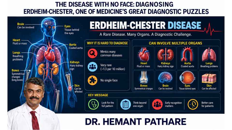
What could possibly connect persistent pain in the long bones of both legs, excessive thirst, tissue appearing around the kidneys, fluid around the heart and an unusual layer of tissue encasing the aorta?
At first, very little.
One symptom may take a patient to an orthopaedic surgeon. Another to an endocrinologist. A kidney abnormality may involve a nephrologist. Breathlessness or pericardial fluid may lead to a cardiologist. An abnormal scan may raise concern about cancer, fibrosis, infection or an autoimmune disease.
Each specialist may be seeing a genuine piece of the problem.
The difficulty is recognising that the pieces belong to one disease.
That disease is Erdheim-Chester disease, or ECD – an extraordinarily rare disorder whose ability to imitate unrelated illnesses has made it one of medicine’s more unusual diagnostic puzzles.
A recent U.S. estimate placed its incidence at only around 1.3 new cases per 10 million people.
Rarity alone, however, does not explain why ECD is so difficult to diagnose.
The greater problem is that it has no single face.
A Disease That Can Appear Almost Anywhere
Erdheim-Chester disease belongs to a group of disorders called histiocytoses.
Histiocytes are cells related to the body’s immune system, particularly the macrophage family. Macrophages normally perform useful work: they engulf damaged material, participate in inflammation and help coordinate immune responses.
In ECD, abnormal histiocytic cells accumulate in tissues where they do not belong. The surrounding inflammatory reaction and fibrosis can gradually interfere with the normal function of those tissues and organs.
The disease was first described by William Chester and Jakob Erdheim in 1930. For much of the decades that followed, even its biological identity was uncertain. Was it primarily an inflammatory disorder? An immune disease? An unusual proliferation of histiocytes?
Modern molecular biology eventually changed that understanding.
ECD is now recognised as a clonal histiocytic neoplasm – an abnormal population of cells driven in most patients by acquired molecular changes affecting growth-signalling pathways.
But knowing what ECD is biologically does not necessarily make the first diagnosis easy.
Its manifestations can still look like entirely different diseases.
Why Diagnosis Can Take Years
One of the most revealing studies came from the U.S. National Institutes of Health, where 60 people with ECD underwent detailed evaluation.
The average interval between their first manifestations and eventual diagnosis was 4.2 years.
The diseases considered along the way show just how convincingly ECD can disguise itself.
Lymphoma was considered in many patients. Others were investigated for infection, sarcoidosis, autoimmune disease, vasculitis, IgG4-related disease, bone malignancy, brain tumours and even multiple sclerosis.
None of those possibilities is unreasonable when considered in isolation.
That is precisely the problem.
ECD can affect the skeleton, kidneys, major blood vessels, heart, lungs, pituitary gland, brain, tissues behind the eyes and skin.
A physician examining only one organ may therefore see something that resembles a much more common condition.
The diagnostic breakthrough often comes not from one spectacular finding, but from recognising an unusual combination of findings across different parts of the body.
And ECD has developed some remarkably descriptive radiological fingerprints.
The Clues: Hairy Kidneys and a Coated Aorta
One of the most characteristic places ECD appears is in the long bones of the legs.
Imaging often shows symmetrical osteosclerosis – abnormal hardening or increased density – around the ends and shafts of bones such as the femur and tibia.
In the NIH cohort, bone involvement was detected in 95% of patients, even though not everyone experienced bone pain.
That distinction is important.
A disease may leave a strong signature on imaging without producing an equally strong symptom.
The kidneys provide another clue.
ECD can cause abnormal tissue to accumulate around both kidneys. On CT imaging, this produces an irregular appearance radiologists memorably call the “hairy kidney” sign.
The tissue can eventually surround the urinary collecting system and ureters, sometimes causing obstruction, hydronephrosis and impaired kidney function.
Then there is perhaps the most striking radiological description of all.
ECD tissue can form a circumferential sheath around the aorta and its branches.
Radiologists call this the “coated aorta.”
Neither a hairy kidney nor a coated aorta establishes the diagnosis on its own.
Together with characteristic long-bone abnormalities, however, they begin to transform apparently unrelated findings into a recognisable pattern.
Then the Disease Reaches the Heart
This is where ECD becomes particularly important from a cardiothoracic perspective.
The abnormal tissue does not necessarily remain outside the organs.
It can involve the pericardium – the sac surrounding the heart – leading to thickening or fluid accumulation. In more severe cases, a large pericardial effusion can compress the heart and produce cardiac tamponade.
ECD may also form an infiltrative mass around the right atrium and right atrioventricular groove, sometimes resembling a cardiac tumour on imaging.
The coronary arteries can become surrounded by abnormal tissue.
The aorta and its branches may be encased.
The myocardium itself can occasionally be involved.
Cardiovascular involvement may produce arrhythmia, ischemia, heart failure or haemodynamic compromise – but it can also remain surprisingly silent and be discovered only on CT or cardiac MRI. Current expert recommendations therefore include cardiac imaging when ECD is diagnosed, even when cardiac symptoms are not prominent.
For a surgeon, this creates an interesting diagnostic lesson.
An abnormal structure around a great vessel or the right side of the heart may look like a problem belonging entirely to cardiac surgery or oncology.
But the anatomy may be only one visible expression of a systemic disease.
The correct question is therefore not always:
“What is this mass?”
Sometimes it is:
“What else is happening elsewhere in this patient?”
That change in perspective can completely alter the diagnosis.
The Lungs Can Provide Another Piece to the Jigsaw Puzzle
The lungs provide another example of ECD’s ability to confuse.
Imaging may show thickening between the small structural units of the lung, ground-glass changes, pleural abnormalities or fluid around the lungs. Pulmonary function can demonstrate a restrictive pattern.
Yet some patients with radiological lung involvement have few respiratory symptoms.
Consensus recommendations estimate that pulmonary abnormalities can be detected on imaging in roughly half of patients.
Again, the disease refuses to behave like a disorder confined neatly to one specialty.
Bone disease may coexist with lung infiltration.
A pericardial effusion may coexist with tissue around the kidneys.
A patient with excessive thirst may have an abnormality involving the pituitary gland rather than ordinary diabetes mellitus.
That last distinction is particularly important.
ECD can cause central diabetes insipidus, a condition in which the body cannot properly regulate water because of disturbance of the hormone vasopressin. It is entirely different from the high-blood-sugar disease usually meant by “diabetes.”
In some patients, this endocrine problem can precede the eventual ECD diagnosis by years.
The clues may therefore appear one by one, separated not only by medical specialties, but by time.
The Biopsy Is Important – But Even the Biopsy May Need Context
Once ECD is suspected, imaging alone is generally not enough.
A tissue biopsy is strongly recommended.
Under the microscope, pathologists look for characteristic histiocytes, often described as foamy because their cytoplasm has a lipid-rich appearance, together with varying degrees of inflammation and fibrosis.
Immunohistochemistry helps establish their identity. Typical ECD histiocytes express markers such as CD68 and CD163while lacking markers such as CD1a that would suggest some other forms of histiocytosis.
Yet even pathology can be deceptive.
Some biopsies contain extensive fibrosis with relatively few characteristic cells. A sample taken from one organ may look less typical than a sample from another.
That is why modern ECD diagnosis is not based on pathology in isolation.
It requires the physician, radiologist, pathologist and increasingly the molecular laboratory to put the evidence together:
Where are the lesions?
What pattern do they form?
What cells are present?
And what molecular alteration is driving them?
It is medicine functioning as pattern recognition rather than a single-test answer.
The Mutation That Changed the Disease
One discovery transformed ECD more than almost anything that came before it.
Researchers found that many patients carry an acquired mutation called BRAF V600E.
BRAF is part of the MAPK pathway, a signalling system that helps cells decide when to grow, divide and survive.
The easiest way to understand the pathway is as a chain of molecular switches.
Normally, a signal turns the pathway on when needed and then allows it to switch off.
Mutations such as BRAF V600E can leave parts of that signalling system abnormally activated.
BRAF V600E is found in roughly half of ECD patients in major cohorts, while alterations activating the wider MAPK pathway are identified in a large majority of patients.
That finding did more than improve classification.
It created a treatment target.
Drugs that inhibit mutant BRAF, such as vemurafenib, can directly interrupt the abnormal signalling driving BRAF-mutated ECD. Patients without a BRAF V600E mutation may harbour abnormalities elsewhere in the pathway that can make MEK inhibitors useful.
A disorder once approached largely through anti-inflammatory, immune-modulating or chemotherapy-based treatments entered the era of molecularly targeted medicine.
This is one of the most remarkable aspects of the ECD story.
Medicine did not merely discover a better drug.
It discovered that it had misunderstood the fundamental nature of the disease.
Could Nanoparticles Help Find It Earlier?
Now another technology is attracting attention.
In 2025, researchers proposed using nanoparticle-enhanced diagnostic methods to improve recognition of ECD.
One particularly interesting avenue involves a nanoparticle called Macrin.
Macrin is designed to accumulate preferentially in macrophages. When labelled with the radioactive isotope copper-64, ^64Cu-Macrin can be detected using PET imaging.
Why might that matter in ECD?
Because the lesions are rich in abnormal cells belonging to the macrophage/histiocyte lineage.
In principle, an imaging agent attracted to macrophage-rich tissue could provide a different way of visualising disease activity – potentially helping identify lesions or choose areas for biopsy.
A first-in-human study published recently evaluated ^64Cu-Macrin in 10 participants, including healthy volunteers and patients with inflammatory or malignant conditions, and explored its safety and ability to image macrophage-rich tissue.
Other nanoparticle approaches being explored include highly sensitive methods for detecting molecular biomarkers and improving analysis of small cell samples. A 2025 publication specifically discussed how such technologies might eventually help overcome some of ECD’s diagnostic limitations.
But there is an important distinction between promise and proof.
Nanoparticle imaging is not currently an established diagnostic test for Erdheim-Chester disease.
The available human Macrin study was extremely small and was not designed to prove that the technique can diagnose ECD.
For now, ECD diagnosis still depends on recognising the clinical and radiological pattern, obtaining appropriate tissue and performing molecular testing.
Nanotechnology represents a fascinating next possibility, not yet the answer.
And that may actually make the story more interesting.
The Disease Did Not Change. Medicine Learned How to See It.
For most of the last century, Erdheim-Chester disease remained difficult partly because medicine was looking for a disease with one recognisable appearance.
ECD does not provide one.
It may look like bone disease in one patient, retroperitoneal fibrosis in another, an endocrine disorder in a third, and cardiac or neurological disease in someone else.
Its identity emerges from the connections.
A coated aorta.
Hairy kidneys.
Symmetrical long-bone lesions.
A right atrial pseudotumour.
Pituitary disease.
Foamy histiocytes.
A MAPK-pathway mutation.
No single clue necessarily tells the whole story.
Together, they can.
And that is perhaps the broader lesson Erdheim-Chester disease offers medicine.
The hardest diagnosis is not always the disease with no clues.
Sometimes it is the disease with many clues that appear to have nothing to do with one another.
Molecular medicine has already made those connections much easier to recognise and has given clinicians treatments directed at the biological machinery of the disease itself.
Nanoparticle imaging may eventually allow medicine to see those abnormal cells in another way.
But the central diagnostic principle will remain much older than either genomics or nanotechnology:
when several organs seem to be telling different stories, sometimes the real task is to discover whether they are all chapters of the same disease.
What do you think is the greater challenge in diagnosing rare diseases – the absence of clues, or recognising when seemingly unrelated clues actually belong together?
